STUDY OF BIOMATERIALS AND SURFACES IN TREATMENTS WITH DENTAL IMPLANTS
|
Description |

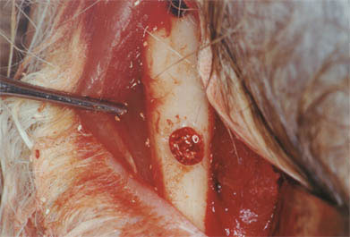
Rabbit tibia in which a bone cavity has been made for filling with biomaterial.
Assessment of the interface between bone tissue and different biomaterials, as well as modification of implant surfaces to enable a faster and more efficient response in implant treatments.
|
How does it work |

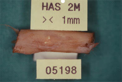
Bone piece of study where a correct integration of the studied biomaterial is observed macroscopically.
Experimental animals (New Zealand rabbits, beagle dogs and minipigs) are used. After surgery in the maxillary, tibial, costal or cranial region, bone defects and implant placement are created. After a period of rest the corresponding samples are obtained that are included in methacrylate, proceeding to histological study by histomorphometry and scanning electron microscopy.
|
Advantages |
It makes it possible to carry out comparative studies between different biomaterials and implant surfaces and gives us precise information about their efficacy.

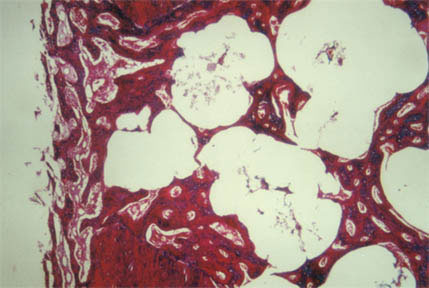
Microscopy study showing biomaterial granules immersed in bone tissue.
|
Where has it been developed |
The studies are done in the Department of Stomatology III (Medicine and Bucofacial Surgery) of the Complutense University of Madrid, in the Service of Experimental Surgery of the Gómez Ulla Military Hospital, and the processing of the samples in the Faculty of Veterinary Medicine of Lugo Veterinarian ros Codina).
|
And also |
Studies of different types of implants and biomaterials patented and commercialized by different companies, being able also:
- Adapt the technology to the specific problems of the client.
- Conduct technical feasibility studies for a specific application.
- Training for the use of the technology in question, etc.
|
Contact |
|
© Office for the Transfer of Research Results – UCM |
|
PDF Downloads |
|
Classification |
|
Responsible Researcher |
José María Martínez González: jmargo@odon.ucm.es
Department: Department of Dental Clinical Specialties
Faculty: Odontology


