DEVICE AND METHOD FOR THE INCREASE OF THE RETINAL REFLEX IN THE PUPIL
|
Description |
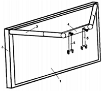
A device has been developed that allows the increase of the retinal reflex when it is not possible to achieve complete neutralization of the shadows in the pupil or in the pupils of patients with miosis. The invention consists of a Fresnel-type lens framed in a rigid frame to which several bars are attached to be able to link the device to the optometric apparatus, easily and comfortably. The device allows the neutralization of the shadows in the retina and the correct measurement of the retinoscopy.
|
How does it work |
Scheme: 1. Fresnel type A magnification lens = 60 cm2 2. Frame.3. Extendable cylindrical bars 4. Flat bar 5. Crimping devices to the optometric instrument.
Under usual conditions, the diameter of the average pupil is 4 mm. With this size, any shadow produced in the retinal reflex that occurs in the pupil can be observed by its direct illumination. When variable pupillary reflexes or a very bright reflex appear, the professional is unable to ensure neutralization. In the first case, they are effects produced by the accommodation or by the existence of irregularities in the cornea that do not allow observation.
In the second case, these are problems due to miosis or pupils with diameters lower than the average value. These pupils are very common in older people with glare problems and try to look for shadows in so little space and neutralizing them becomes impossible.
This new device allows the measurement of retinoscopy with miotic pupils
The device can be removed and put with comfort and speed, being compatible and, therefore, universal with all kinds of measuring instruments. The magnification sheet can be changed away from the eye to vary the size of the pupil at the whim of the observer.
|
Advantages |
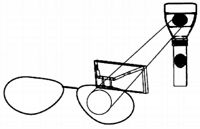
Operation of the device. It is observed how the light passes through the magnifying lens and is transmitted to the pupil.
The major drawbacks of the measurement of retinoscopy are the intervention of accommodation and changes in pupillary diameter. With a device of this type, at a reasonable price for universal use, the observation of an important sector of the population is easily solved.
|
Where has it been developed? |
The design, protected by national patent with previous examination since 2012, and its prototype, have been developed in the Faculty of Optics and Optometry.
|
Contact |
|
© Office for the Transfer of Research Results – UCM |
|
PDF Downloads |
|
Classification |
|
Responsible Researcher |
Ricardo Bernárdez Vilaboa: rbvoptom@ucm.es
Department: Optometry and Vision
Faculty: Optics and Optometry


