BRAIN MAPPING AND FUNCTIONAL NEUROIMAGING (PET)
|
Description |
The Center includes two main laboratories: 1) human brain mapping with high resolution electroencephalography (EEG) for cognitive studies, and 2) Molecular imaging with PET (Positron Emission Topography) instrumentation in animal experimental models.

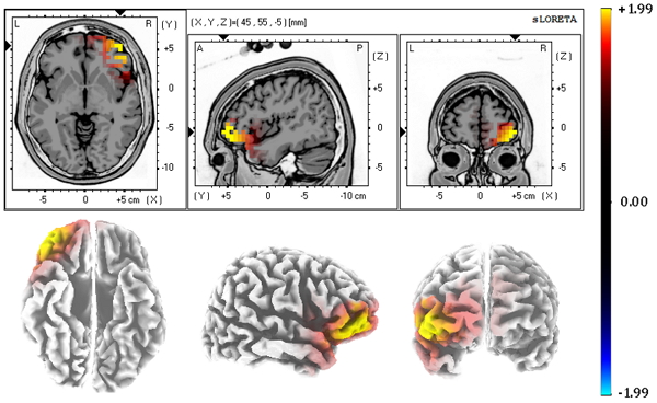
Image of the location of neural origin, by applying the sLORETA algorithm, of the EEG activity recorded in the scalp
|
How does it work |
EEG
The technique uses the electrical brain activity from volunteer participants while they perform several tasks. The participant receives certain sensory stimulation that evokes electrical changes over time in various parts of the brain. The technique incorporates the Brain Vision software to register the echoes of such electrical activity at the surface of the human scalp, non-invasively.
Two recording sets are available (one with 32 and the other with 64 head electrodes). Both have the possibility of adding 6 additional external channels, two of them as a reference site and four more for recording the electro-oculogram. Data recorded at the scalp are used to estimate the potential generating brain sources. That is, we are able to determine three-dimensional coordinates of cortical brain areas that became activated during the task.
PET
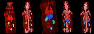
The PET/CT technique for small animals consists of the injection of a radiotracer to which a positron emission isotope has been incorporated. Depending on the tracer properties it will differentially accumulate in various body sections. Its detection relies on the emission of a positron by the isotope which at the encounter of a neighboring electron suffers an annihilation radiation. During that process, two anti-parallel high-energy gamma rays are generated and become detected by the PET ring. A subsequent reconstruction process allows to obtain three-dimensional images showing the distribution of the radiotracer. Most commonly, the use of 18F-FDG, offers a map of metabolic activity. Studies using this technique are relevant to multiple fields: physiology, pharmacology, oncology, neurology, new drug development, experimental models, etc. Structural information recorded by the TAC module complements data acquisition. . Most commonly, the use of 18F-FDG, offers a map of metabolic activity. Studies using this technique are relevant to multiple fields: physiology, pharmacology, oncology, neurology, new drug development, experimental models, etc. Structural information recorded by the TAC module complements data acquisition.
|
Advantages |
The application of these techniques offers multiple advantages:
EEG
- It is a non-invasive technique, suited for cognitive ERP studies.
- It has an excellent temporal resolution: able to detect changes in the order of milliseconds.
PET/CT
- High sensibility Molecular Imaging in animal disease models.
- Minimally invasive technique, with the injection of a molecular radiotracer.
- Longitudinal studies are possible within the same animal in order to assess pathological and treatment progression.
- High resolution TAC images are possible to obtain, allowing a very precise anatomical localization.
- The CT technique is also suited for inanimate objects: bone tissue, prosthesis, rocks, various materials, etc.

PET-CT image
|
Where has it been developed |
These techniques have been developed in the Centro de Cartografía Cerebral of the Complutense University of Madrid. This center offers support to the research activity of the UCM University. It also provides support to other public and private centers throughout various agreements and conventions. The Center holds the IS- 9001 quality certification.
|
And also |
EEG, Brain Mapping:
Nowadays this technique has been demanded from researchers and companies in need of cognitive studies. We collaborate with various research groups from the Complutense University and other national and international Universities.
PET/CT Functional Imaging:
The laboratory has a wide expertise offering PET services to research teams at the Complutense and other Universities, to other research centers and other companies that develop studies on preclinical pharmacological substances.
We collaborate with the Instituto Tecnológico PET, a company that has a medical cyclotron for the production of positron-emitting radionuclides, and with chemical synthesis groups in the development of new radiotracers.
The laboratory includes rodents animal house (ES280790000197) and radioactive facility (type 2) recognized and authorized by the Nuclear Security Board (The Spanish Consejo de Seguridad Nuclear).
|
Contact |
|
© Office for the Transfer of Research Results – UCM |
|
PDF Downloads |
|
Classification |
|
Responsible Researcher |
Miguel A. Pozo García: pozo@ucm.es
Multidisciplinary University Institute
Brain Cartography Center


